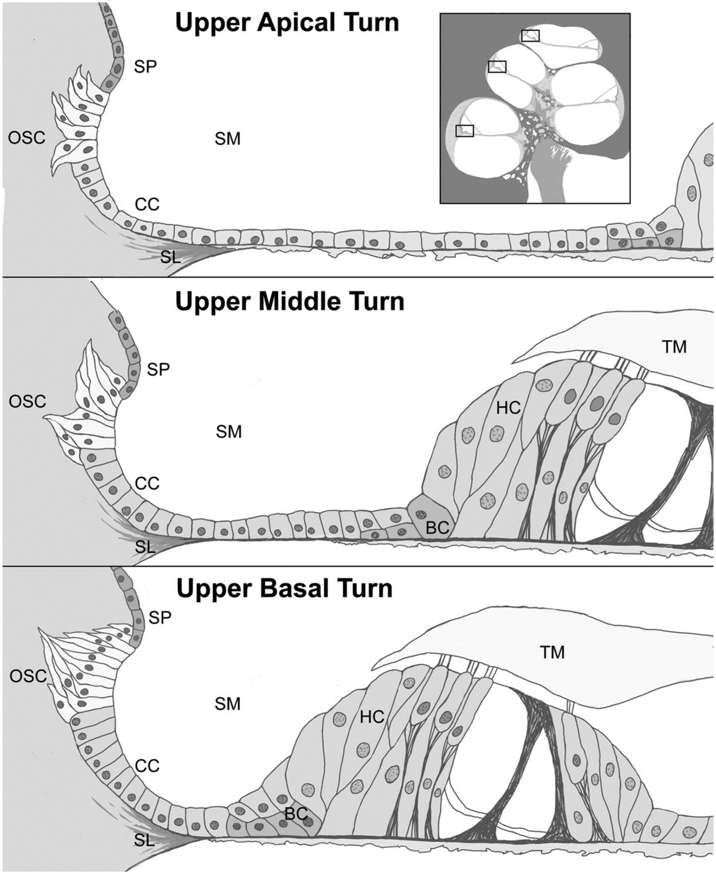
FIG. 3.

Idealized rendering of the cellular structures of the basal, middle, and apical turns of the human cochlea based on light and confocal microscopy findings. Outer sulcus cells extend into the spiral ligament in bundles forming tapering processes which differ between the cochlear turns in morphology. OSC indicates outer sulcus cell; SP, spiral prominence; CC, claudius cell; SL, spiral ligament; BC, Boettcher cell; HC, Hensen’s cell; TM, tectorial membrane; SM, scala media.
