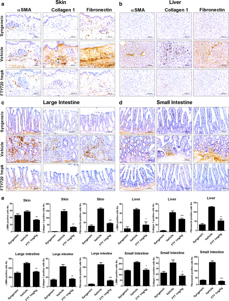
Fig. 6.
FTY720 downregulated the marker proteins of fibrosis in tissues of a murine model of GvHD. a–d Representative examples of immunohistochemical staining for αSMA, Collagen1, and Fibronectin in skin, liver, large and small intestine tissue from mice with GHVD. Positive immunoreactivity appears as a brown color, and tissues are counterstained with blue (original magnification, ×400). e Bars represent the average number of positive cells in the skin, liver and large and small intestine, of each groups. Tissues were collected on day 28 after transplantation

