Fig. 1.
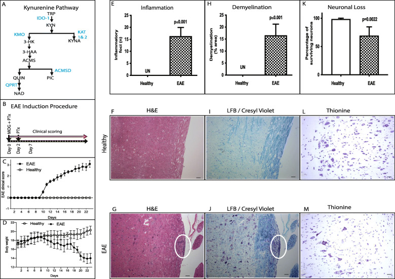
Experimental autoimmune encephalomyelitis (EAE). a Schematic illustration of kynurenine pathway (KP). KP is depicted as metabolites in black, enzymes in blue. b Experimental procedure to induce experimental autoimmune encephalomyelitis (EAE) in C57BL/6 mice with MOG35-55 peptide (myelin oligodendroglial protein) and pertussis toxin (PTx). c Mean daily clinical score in the EAE (n = 18) and healthy mice (n = 15). d Body weight of EAE and healthy mice was determined (mean ± SD). e–i Inflammation, demyelination, and neuronal loss in EAE-induced mice spinal cord. e The inflammatory foci (mean ± SD) counted in H&E stained axonal sections of EAE and healthy control. f and g Representative histological spinal cord sections of healthy (f) and EAE mice (g) stained with H&E. h Demyelination (% area) without inflammation detected by Luxol Fast Blue (LFB)/cresyl violet staining. i and j Representative histological spinal cord sections of healthy (i) and EAE mice (j) stained with LFB/cresyl violet. k Surviving neurons stained for Nissl substances using 0.1% thionine and neurons with well-defined nucleolus were counted. l and m Representative histological spinal cord sections of healthy (l) and EAE mice (m) stained with thionine for surviving neurons; white oval shows inflammatory foci (l, n = 6) and demyelinated area (m, n = 6). Scale bar 10 μm (f, g, i, and j), 25 um (l and m). IDO-1, indoleamine 2,3-dioxygenase; KAT, kynurenine amino transferase; KMO, kynurenine monooxygenase; KYNU, kynureninase; 3HAO, 3-hydroxyanthranilate 3,4- dioxygenase; ACMSD, 2-amino-3-carboxymuconate-semialdehyde decarboxylase; QPRT, quinolinate phosphoribosyl transferase

