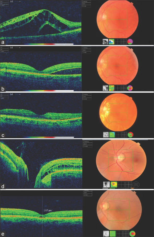
Figure 1.

(a) Optical coherence tomography scan of the left eye showing a serous macular detachment with subretinal fluid. (b) Optical coherence tomography scan carried out at a 2-month postoperative follow-up. Central fovea is flat, whereas the subretinal fluid in the macular area appears to be substantially reduced. (c) Image from optical coherence tomography scan 6 months postoperatively. The fovea is clear of fluid, and there is a significant regression of the subretinal fluid. (d) Optical coherence tomography scan showing optic disc pit maculopathy with intraretinal fluid and cystic spaces. (e) Resolution of maculopathy after focal laser application. Image on a 2-year follow-up
