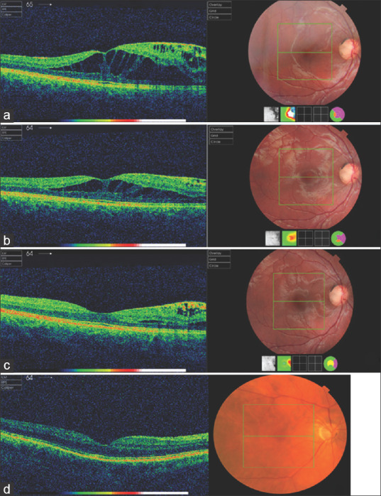
Figure 2.

(a) Optical coherence tomography scan of the right eye showing an optic disc pit in the temporal aspect of the optic nerve head with adjacent intraretinal fluid. (b) Optical coherence tomography scan, 3 months postoperatively, indicative of gradual improvement of the intraretinal fluid. (c) Last follow-up appointment. The intraretinal fluid at the optical coherence tomography scan has almost completely resolved. (d) Optical coherence tomography scan of the right eye showing optic disc pit without any related foveal abnormalities
