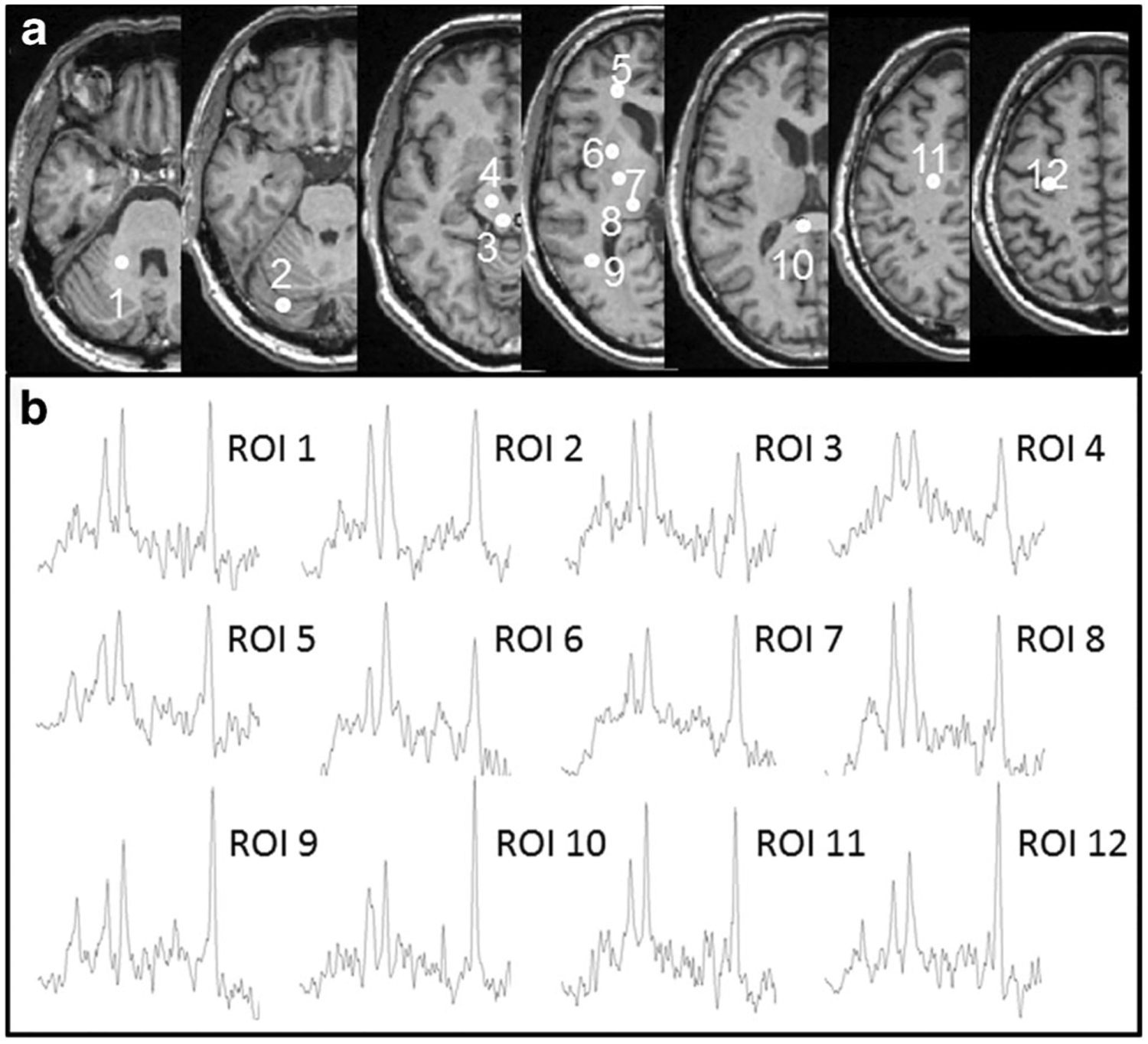
Fig. 2.

a Locations of selected regions of interest (ROIs) in the right brain hemisphere displayed as white filled circles on T1 weighted images of a volunteer (male, 52 years). The consecutive numbers represent the ROIs located in cerebellar white matter at level of middle cerebellar peduncle (Cblwm, 1), cerebellar anterior lobe at level of pons (Cbla, 2), midbrain tegmentum (MDd, 3) and cerebral peduncle (MDv, 4), frontal white matter (fWM, 5), putamen (6), posterior limb of the internal capsule (iCap, 7), thalamus (8), parietal white matter (pWM, 9), splenium of the corpus callosum (sCC, 10), centrum semiovale (CSO, 11) and hand motor area (HN, 12). b Example MR spectra in each of the ROIs in cerebellum and cerebrum as indicated, derived from whole-brain magnetic resonance spectroscopic imaging data
