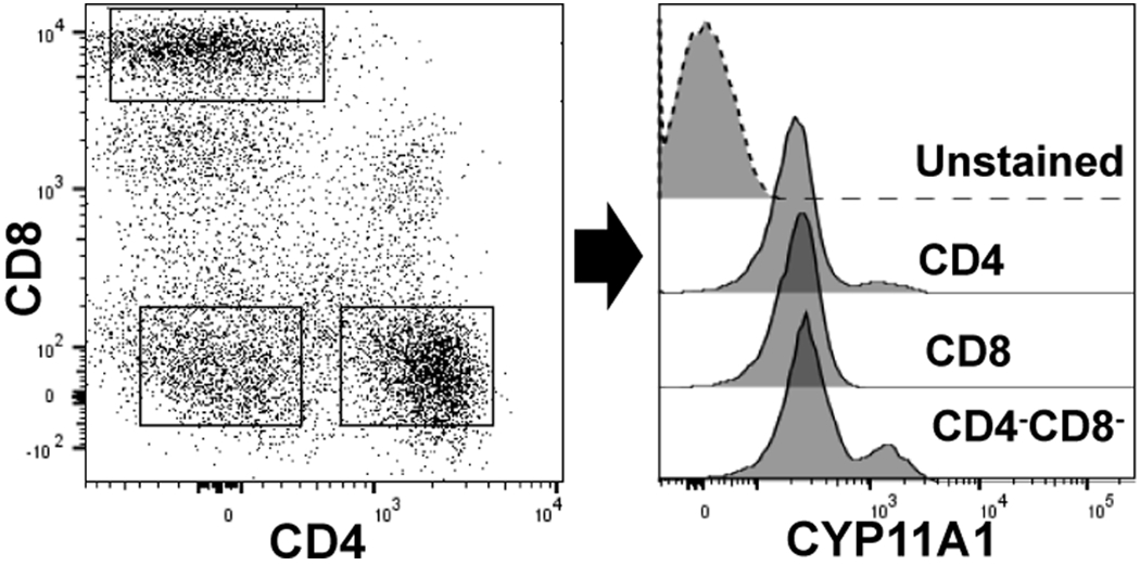
Figure 3:

CYP11A1 expression in human peripheral blood mononuclear cells (PBMCs).The left dot plot shows CD4, CD8 T cells, and CD4−CD8− cells in PBMCs. The right histogram shows expression of CYP11A1 in gated CD4 cells, CD8 T cells, and CD4−CD8− cell populations verses the unstained PBMC. The blood was obtained from a healthy volunteer (IRB 160426001) and processed as described previously250. Intracellular staining for CYP11A1 (Cell signaling technology; Danvers, MA, USA) was performed in cells fixed with paraformaldehyde and permeabilized in methanol containing buffer 250, 251. Anti- Cyp11A1 was conjugated to APC-Cy7 (Abcam; Cambridge, UK) as per manufacturers protocol before use. Stained cells were analyzed using a BD-FACS Symphony flow cytometer (BD Biosciences, San Jose, CA). Data are representative of three independent experiments utilizing different donors.
