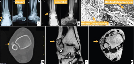
Figure 1.

Case 1, a34-year-old female with an exophytic growth (a) with progression of lesion after 10 months (b). Biopsy showing histopathological signs of bizarre parosteal osteochondromatous proliferation (c). Computed tomography axial section showing discontinuous outgrowth from fibula (d). Magnetic resonance imaging scans showing a focal mature osteocartilaginous mass on lateral surface of fibula showing heterogenicity (e and f).
