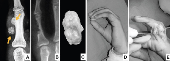
Figure 4.

Case 2, a 21-year-old male with (a) pre-operative X-ray showing an ossified nodule on the surface of the phalanx (arrow) and a separate ossified mass in the soft tissue. (b) Post-operative X-ray showing the excised cortex. (c) Excised specimen as seen from the surface. The bony cortex is on the far side and not visible in this picture. (d) Clinical photo at presentation. Note the swelling and scar of the previous surgery. (e) Intraoperative photograph showing the lesion on the phalanx.
