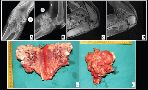
Figure 5.

Case 3, a31-year-old male with (a and b) pre-operative X-ray, the X-ray showed large calcific lesion arising from the upper end of tibia and likely from fibula. The calcification was chunky and irregular in nature and appeared to be a secondary sarcomatous change in a case of long existing osteochondroma and magnetic resonance imaging (c and d) suggested of an irregular osteochondromatous lesion extending toward the posterior neuromuscular bundle but not invading it. (e and f) Excised specimen consisting of tibia and fibula.
