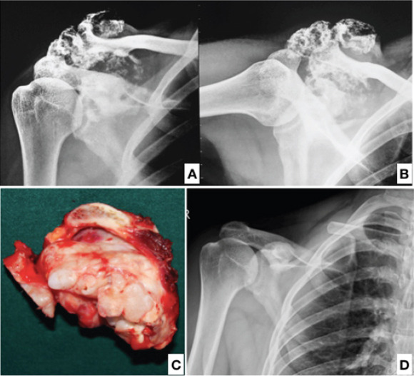
Figure 7.

Case 4, a 26-year-old male with X-rays showing a large osseous mass over the lateral end of the clavicle (a and b). Operative specimen of the excised lateral end of clavicle showing a lobulated osseous exophytic growth (c). The patient on a yearly follow-up showed no signs of recurrence and had good clinical function (d).
