
Figure 3.
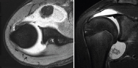
Fat-suppressed T2-weighted axial (a) and coronal oblique (b) magnetic resonance images of the right shoulder show a large mass lesion in the subscapular bursa and loose bodies in the axillary pouch.
Official websites use .gov
A
.gov website belongs to an official
government organization in the United States.
Secure .gov websites use HTTPS
A lock (
) or https:// means you've safely
connected to the .gov website. Share sensitive
information only on official, secure websites.
Fat-suppressed T2-weighted axial (a) and coronal oblique (b) magnetic resonance images of the right shoulder show a large mass lesion in the subscapular bursa and loose bodies in the axillary pouch.