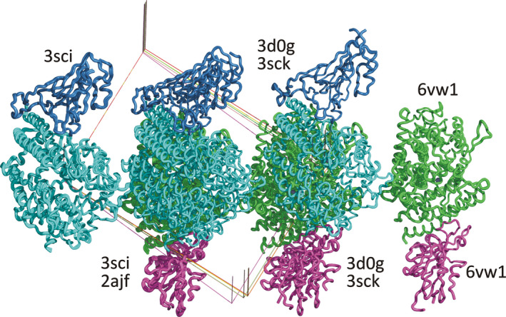
Fig. 2.

Lack of uniformity in presentation of structures in the PDB. Placement of selected models of the complexes of the receptor‐binding domains of CoV and CoV‐2 spike proteins with the proteolytic domain of ACE2 in isomorphous unit cells in space group P21. The individual structures are identified by their PDB codes. The chains of ACE2 are colored cyan/green, and the RBDs of the spike proteins blue/magenta for the two complexes present in each asymmetric unit. Figure generated with pymol.
