Fig. 4.
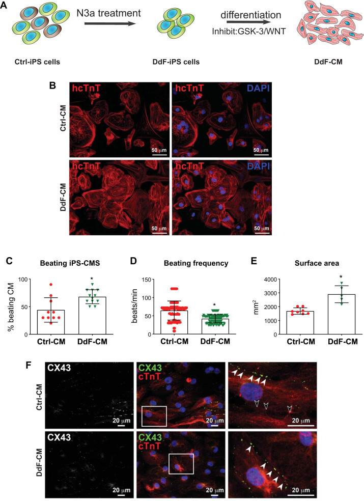
DNA-damage-free induced pluripotent stem cells (DdF-iPS cells) show enhanced cardiomyocyte (CM) differentiation. A: schematic of DdF-iPS cell selection and differentiation. B: immunolabeling of human-specific cardiac troponin T (hcTnT; red; all panels) and nuclei counterstained with DAPI (blue; right) in control (Ctrl) CM (top) and DdF-CM (bottom). C: the fraction of beating CMs generated by differentiating Ctrl-iPS and DdF-iPS cells. n = 3 (total 10–12 fields analyzed in each group) in all cases. Significance was analyzed by t-test. Data are means ± SD. *P < 0.05 vs. Ctrl-CM. D: frequency of beating by Ctrl-CM (n = 3, total 100 CMs) and DdF-CM (n = 3, total 120 CMs). t-test was performed. Data are means ± SD. *P < 0.05 vs. Ctrl-CM. E: the average surface area of Ctrl-CM and DdF-CM. n = 3 (total 5–8 fields analyzed in each group) in all cases. t-test was performed. Data are means ± SD. *P < 0.05 vs. Ctrl-CM. F: confocal imaging of the immunofluorescence-labeled gap junction protein connexin 43 (CX43) and cardiac troponin T in Ctrl-CM and DdF-CM (white arrowheads, CX43 in cell-to-cell junction; black arrowheads, absence of CX43 in cell-to-cell junction). N3a, nutlin-3a.

