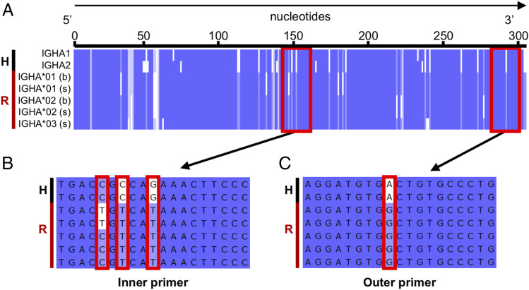
FIGURE 5.
Comparison of human and rhesus IgHA reference sequences and illustration of human 10× primer target regions. A black H and red R, respectively, highlight human and rhesus sequences on the left side of each panel. (A) Multiple sequence alignment of putative human IgHA1 (J00220) and IgHA2 (J00221) consensus domain sequences with rhesus consensus cDNA coding sequences recovered (IgHA*01, IgHA*02, and IgHA*03) in either secreted (s) or membrane-bound (b) form. The target sites of human 10× primers are denoted by red boxes. (B and C) Zoom in of the inner and outer primer target sites, where mismatches between the primer and rhesus consensus sequence(s) are denoted by red boxes.

