Figure 2.
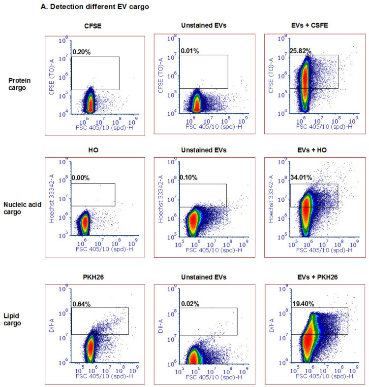
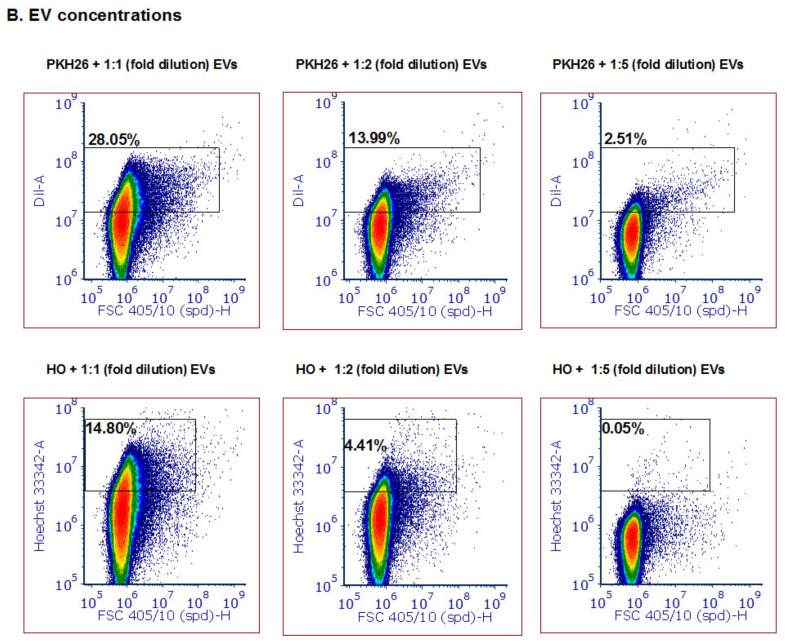
Detection of different types and amounts of Pf-derived EV cargo using the ZE5 analyzer. (A) Pf-derived EVs were stained using three different dyes to visualize different types of cargo components: proteins with the CFSE marker (top row), nucleic acids with the HO marker (middle row), and lipids with the PKH26 marker (bottom row). As controls, solutions containing free dye or unstained EVs were used. (B) Detection of different Pf-derived EV concentrations was achieved by diluting the EVs with the same dye (PKH26 and HO) amount. On the left: high concentration (8 × 10¹¹ par/mL); in the center and on the right: lower concentrations of EVs.


