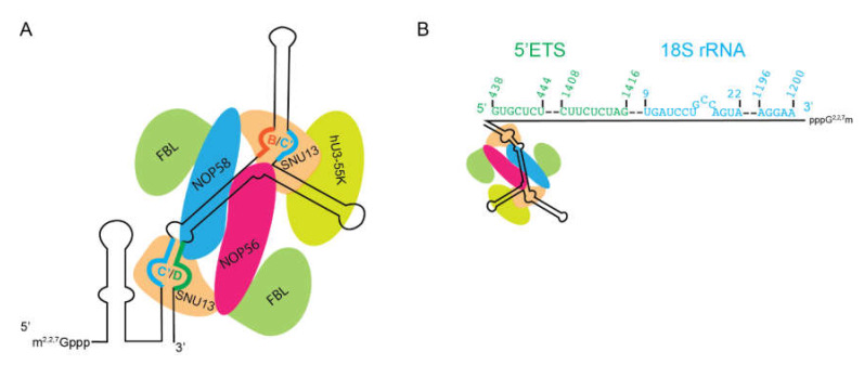
Figure 5.
U3 snoRNP. (A) Secondary structure of the U3 snoRNA with protein components. Note that SNU13 still binds k-turns, here formed by Box C′ (blue) and Box D (green) and Box C′ (blue) and Box B (Red). The altered structure of the U3 snoRNA results in FBL not being positioned for 2′-O- methylation. (B) The 5′ extension forms multiple base-pairs with the pre-rRNA. Two different stretches in the 5′ETS (green) and the 18S rRNA (blue) are bound. Dashed lines indicate looped-out stretches of rRNA. Numbers indicate position in the 47S rRNA (green) or 18S rRNA (blue). Note that this schematic does not fully represent looping out. Secondary structure modified from predictions on snoRNABase [95].

