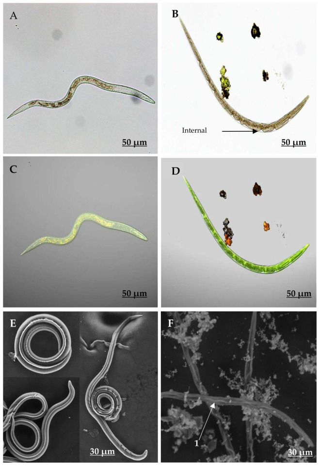
Figure 4.
Micrographs taken with AxioCam (A,B), Confocal Laser Scanning Microscopy (CLSM, C,D) and Environmental Scanning Electron Microscopy (ESEM, E,F) showing the aspect of Haemonchus contortus larvae exposed (B,D,F) and non-exposed to isorhamnetin (A,C,E). F1 shows the wave formations at the lateral nervous cord.

