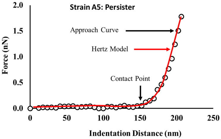
Figure 11.
A representative indentation-force profile obtained on one of the A5 persister cells. The location of the contact point (CP) was obtained manually from the AFM Nanoscope Analysis 1.5 software (Bruker, Camarillo, CA, USA) and indicated by the downward black arrow. The force–indentation data were obtained from a persister cell exposed to a 1000 μg/mL of ampicillin for 25 h.

