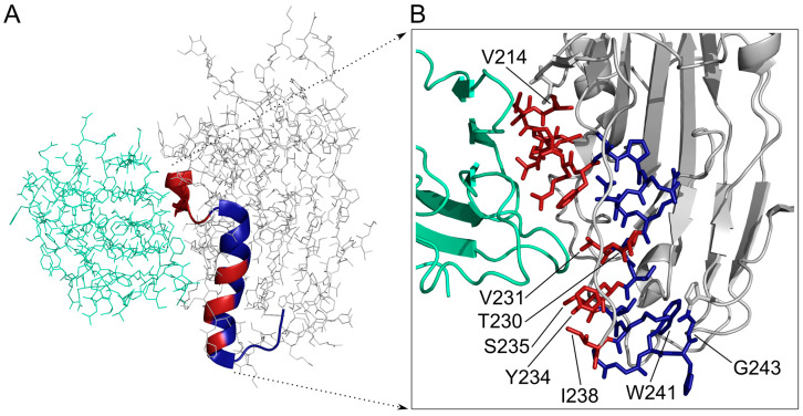
Figure 9.
3D structure of HSV-1 gD glycoprotein (grey) in complex with nectin-1 (ocean green). (A) the ribbon indicates the region covered by Set III peptides, red parts participate in the HSV-1 gD – nectin-1 interaction. (B) Details of the binding surface with HSV-1 gD 214–243 region are in stick representation, red residues are in contact with nectin-1. After the strongly binding 214–223 segment (red) the non-contacting 224IPENQR229 segment can be seen (blue), then 230TVAVYSLKI238 helical region with alternating contacting and non-contacting residues. Segment 240GWHG243 (blue) is folding back on the helical region.

