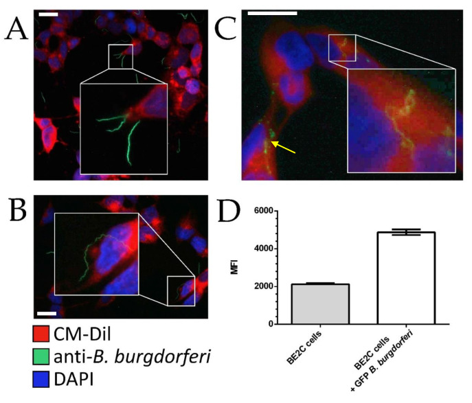
Figure 1.
Spatiotemporal interactions between BE2C cells and B. burgdorferi. BE2C cell co-culturing at 30 min (A), 2 h (B), and 24 h (C) timepoints were visualized via Figure 2. C cell boundaries while polyclonal anti-B. burgdorferi was used to visualize B. burgdorferi. (D) After 24 h co-culture with green fluorescent protein (GFP)-labeled B. burgdorferi, BE2C cells were analyzed by flow cytometry to assess GFP fluorescence, indicating cell–cell interactions. All images were taken at 400× magnification. Scale bar is 25 µm. MFI, mean fluorescence intensity. DAPI, 4’,6-diamidino-2-phenylindole.

