Figure 4.
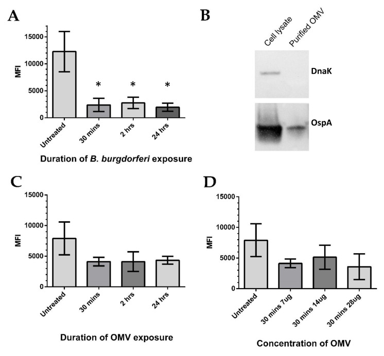
B. burgdorferi and outer membrane vesicles (OMVs) decrease BE2C cell mitochondrial superoxide levels. (A) Flow cytometry was used to measure the mean fluorescence intensity (MFI) of the mitochondrial superoxide indicator, Mitosox, in BE2C cells. Increasing co-culture times with B. burgdorferi results in BE2C cell superoxide reduction. (B) B. burgdorferi OMVs were isolated via ultracentrifugation and purity verified by observing an absence of the intracellular protein DnaK from OMV isolates and presence of membrane-specific OspA. (C) BE2C cell superoxide decreased after treatment with OMVs, but was not statistically significantly compared with the untreated conditions. Increasing OMV exposure durations did not produce a proportional superoxide decrease. (D) BE2C cell superoxide levels decreased in comparison with untreated cells, but were not statistically significant compared with the untreated conditions in response to B. burgdorferi OMVs at varying OMV amounts. An unpaired Mann–Whitney test was used to determine significance between treated and untreated conditions (* significant at p < 0.05). Error bars represent SEM of six experiments.

