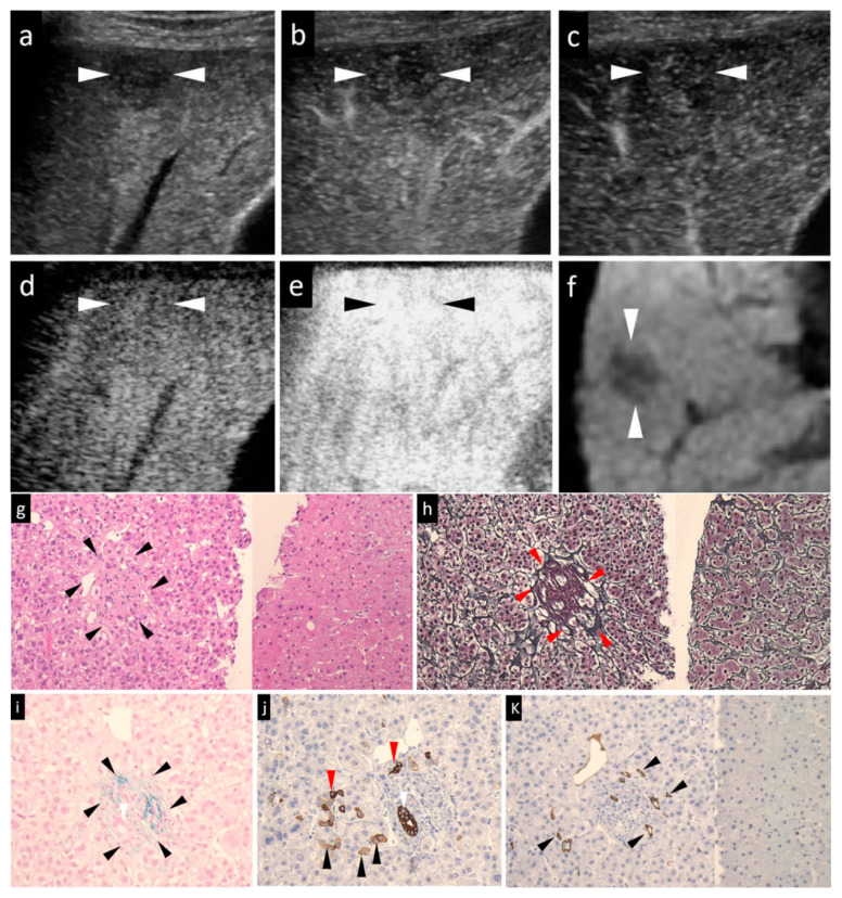
Figure 1.
A case of early HCC (maximum diameter, 15 mm) in segment V with the typical imaging patterns and histological staining. The patient was a 65-year-old male. (a) Grayscale US showed a slightly hypoechoic lesion in segment V, near the surface of the liver. Neither the halo nor the mosaic sign was positive. (b,c) Arterial-phase Sonazoid CEUS image obtained using low-mechanical index (MI) harmonic imaging showed slight transient hypovascularity (b) and the lesion was seen as an isovascular lesion in the subsequent portal-phase image (c). (d) Post-vascular phase CEUS image obtained with low-MI contrast imaging showed an isoechoic tumor. (e) After the imaging mode was switched to high-MI contrast imaging, the post-vascular phase showed an isoechoic tumor. (f) The lesion was seen as a hypointensity in segment V in the hepatobiliary-phase image of Gadolinium ethoxybenzyl diethylenetriamine pentaacetic acid magnetic resonance imaging (Gd-EOB-DTPA MRI). (g) Hematoxylin–eosin (HE) staining. As compared to the non-tumor area (right side), early HCC (left side) showed the cancer area (arrowheads) has a slight cellular atypia, a mild increase in the nuclear-cytoplasmic ratio, and hypercellularity. The trabeculae were arranged clearly. (h) Silver staining of the tumor area (left side) and non-tumor area (right side). In early HCC, the reticular fibers were clear and evenly distributed, much resembling the findings in the non-tumor area. Red arrowheads indicated portal tracts. (i) Victoria blue staining of the tumor area showed elastic fibers surrounding the portal tract in blue (arrowheads). Positive stromal (portal tract) invasion, namely, cancer cells within the portal tract, were compatible with the diagnosis of early HCC. (j) Cytokeratin 7 staining of the tumor area. No ductular reaction could be seen in this figure. The progenitor cells were small cells with dense staining of the cytoplasm (red arrowheads). Black arrowheads indicated intermediate hepatocytes with faint staining of the cytoplasm. The white arrowheads in (i) and (j) indicated the presence of bile ducts within the portal tract. (k) CD34 staining. Focal expression of CD 34 (arrowheads) was seen in the cancer area (left side), which was compatible with the diagnosis of early HCC. Absence of CD34 expression in non-tumor area (right side) suggested the absence of any neovascularization in the non-tumor adjacent hepatic tissue. The arrowheads seen in images (a–f) indicated the margin of the lesion.

