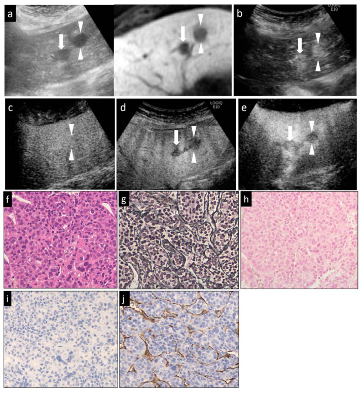
Figure 3.
A hypervascular HCC lesion (maximum diameter, 16 mm) in segment III in a 58-year-old male patient with underlying chronic C hepatitis. (a) Fusion images combining grayscale US (left side) and the hepatobiliary-phase image of Gd-EOB-DTPA MRI as reference (right side) on a single screen. The hepatobiliary-phase image of Gd-EOB-DTPA MRI showed a significant hypointense area in segment III (arrowheads). The target lesion appeared as a well-defined hypoechoic lesion on the grayscale US image. The grayscale US image showed a homogeneous hyperechoic lesion without a surrounding halo. (b) Arterial-phase Sonazoid CEUS images obtained using low-MI harmonic imaging showed a significantly hypervascular area. (c) The lesion appeared isovascular in the portal-phase image, (d) and as a hypoechoic area in the post-vascular image (washout). (e) After the imaging mode was switched to high-MI contrast imaging, the lesion appeared as a clear defect in the post-vascular phase. (f) Hematoxylin–eosin (HE) staining showed obvious cancer cell and nuclear atypia, including hypercellularity, increased nuclear-cytoplasmic ratio and larger, deformed nuclei. (g) Silver staining showed that the reticulin formation was slightly unclear in some portions. (h) Victoria blue staining showed no stromal invasion of cancer cells. (i) Cytokeratin 7 staining showed no expression of the marker in the tumor areas, that was an absence of ductular reaction. (j) Diffuse expression of CD34 in the peripheral areas suggested increased neovascularization resulting from sinusoidal capillarization and a formation of sinusoidal vascular endothelium in HCC. This lesion was histopathologically diagnosed as a moderately diff. HCC. The arrowheads seen in (a–e) indicated the margin of the lesion. Another lesion was also seen in (a,b,d,e) (arrows).

