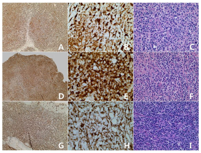
Figure 1.
Hodgkin lymphomas with expression of Indolamine-2,3-dioxygenase (IDO): classic Hodgkin lymphoma, mixed cellularity. (A–C): cervical lymph node; (G–I): palatine tonsil. Some tumor cells and vascular structures show reactivity for IDO protein (A,G): IDO stain, ×40; (B,H): IDO stain, ×400; (C,I): H&E stain, ×400. Nodular lymphocyte predominant Hodgkin lymphoma (D–F): cervical lymph node. Some tumor cells and vascular structures show reactivity for IDO protein (D): IDO stain, ×40; E: IDO stain, ×400; F: H&E stain, ×400).

