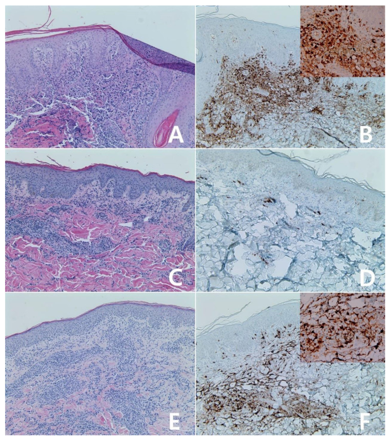
Figure 8.
Immunohistochemistry for IDO in primary cutaneous CD30 positive T-cell proliferative disorder: Lymphomatoid papulosis (A,B): case 1, (C,D): case 2, (A–D) ×100, (A,C): H&E stain. Atypical lymphocytes showed diffuse positivity for IDO protein (B), score 4; Insert: ×400, negative for IDO (D), score 0. One primary cutaneous anaplastic large cell lymphoma was scored 3 (E,F), ×100; Insert: ×400, (E): H&E stain.

