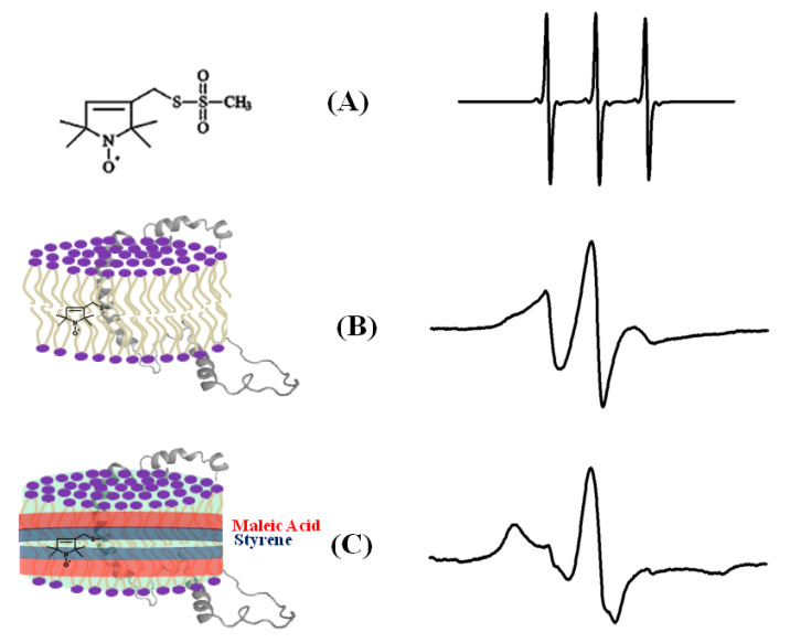
Figure 4.
An illustrative example of the EPR spectra for a spin-labeled membrane protein in different membrane environments. (A) A free MTSL spin label in solution, (B) MTSL spin label on a F56 C-KCNE1 membrane protein in a lipid bilayer, (C) MTSL spin label on a F56C-KCNE1 membrane protein in lipodisq nanoparticles. The CW-EPR spectrum for lipodisq nanoparticle samples also shows a minor peak due to free spin labels. (Adapted from [69] with permission).

