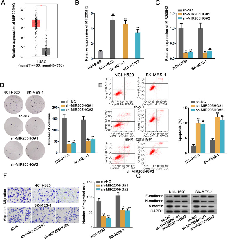
Fig. 1.
Cutting down MIR205HG expression impaired cell proliferation and migration abilities in LUSC. a. GEPIA dataset was applied to examine the relative expression of MIR205HG in LUSC tissues and adjacent normal tissues. b. Carrying out qRT-PCR to determine different expressions of MIR205H in normal lung squamous cell line (BEAS-2B) and LUSC cell lines (NCI-H520, SK-MES-1 and NCI-H1703). c. The relative expression of MIR205H under the condition of down-regulating MIR205H was measured by qRT-PCR analysis. d. Number of colonies was assessed by colony formation assay to detect cell proliferation ability. e. Cell apoptosis rate was estimated by flow cytometry analysis. f. Transwell assay was used to measure cell migration capability by counting number of migrated cells. g. The expressions of the epithelial marker (E-cadherin) and the mesenchymal markers (N-cadherin and Vimentin) were measured by western blot analysis. All data were displayed as the mean ± SD. *P < 0.05, **P < 0.01

