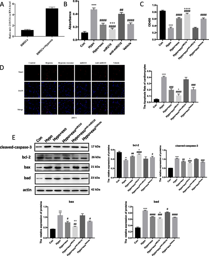
Fig. 4.
Overexpression of miR210 in MSC exosomes strengthens their cardioprotective function in vitro. a Polymerase chain reaction quantification of miR210 in the exosomes obtained from MSCs. Hypoxic cardiomyocytes were treated with exosomes obtained from MSCs that were pre-treated with miR210 (exomiR210), anti-miR210 (exoanti-miR210), or scramble (exoVehicle) for the indicted time and cell injury characterized by LDH release (b), viability by MTT assay (c), and apoptotic programmed cell death by TUNEL assay (d). Red, TUNEL-positive nuclei; blue, DAPI-stained nuclei; green, troponin-positive cardiomyocytes. Scar bar × 200. The percentage of TUNEL-positive cells is shown in the right panels. e Western blot identification of BAD, BAX, BCL-2, and Cleaved-CASPASE-3 in cardiomyocyte lysates after cell incubation with the indicated exosomes ± hypoxia. Quantitative analysis of BAD, BAX, BCL-2, and Cleaved-CASPASE-3 is shown in the lower panel. Each experiment was repeated 3 times. **p < 0.01, ***p < 0.001, ****p < 0.0001 for hypo vs. con. #p < 0.05, ##p < 0.01, ###p < 0.001, ####p < 0.0001 for hypo + exo, hypo + exomiR210, hypo + exoanti-miR210, or hypo + exoVehicle vs. hypo. +p < 0.05, ++p < 0.01, +++p < 0.001, ++++p < 0.0001 for hypo + exomiR210 vs. hypo + exo

