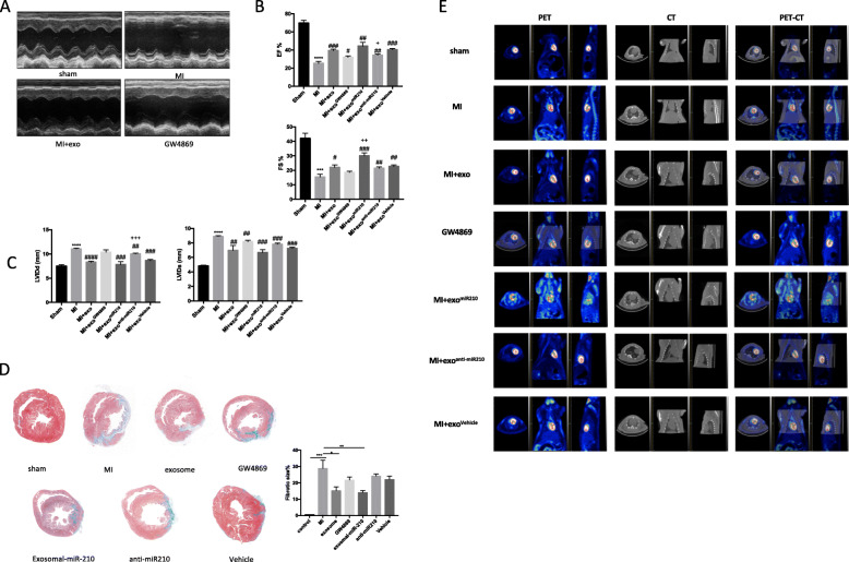
Fig. 6.
Overexpression of miR-210 in MSC exosomes leads to improvement of cardiac function and infarct size in vivo at 1 month after MI. a–c Representative photographs of M-mode echocardiography. Quantitative analysis of echocardiography (N = 4 per group). FS, fractional shortening; EF, ejection fraction; LVIDd, left ventricular internal diameter end-diastolic; LVIDs, left ventricular internal diameter end-systolic. d Masson Trichrome staining of whole heart tissue sections 28 days post-ligation and treatment with PBS, MSC exosomes, exosomes derived from MSCs treated with GW4869, MSCs-exomiR210, MSCs-exoanti-miR210, or MSCs-exoVehicle. Percentage of tissue fibrosis is quantified and presented. N = 4 per group. e Representative photographs of PET-CT of rats from the respective treatment groups. N = 3 per group. ***p < 0.001, ****p < 0.0001 for MI vs. sham. #p < 0.05, ##p < 0.01, ###p < 0.001, ####p < 0.0001 for MI + exosome, GW4869, MSCs-exomiR210, MSCs-exoanti-miR210, MSCs-exoVehicle vs. MI. +p < 0.05, ++p < 0.01, +++p < 0.001 for MSCs-exomiR210, MSCs-exoanti-miR210 vs. MSCs-exoVehicle

