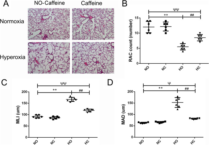
Fig. 3.
Effect of caffeine on pulmonary alveolar simplification in lung. Hyperoxia exposure led to marked alveolar simplification as shown by the HE staining in the images and by the assessment of RAC, MLI and MAD. Caffeine treatment improved the hyperoxia-induced impairment of alveolar growth. a Representative HE staining (light microscopy, × 100) of lung tissue slides from each group. Scale bars = 100 μm. b Semiquantitative pathology determination of RAC in lung tissues. c Semiquantitative pathology determination of MLI in lung tissues. d Semiquantitative pathology determination of MAD in lung tissues. The values are the mean ± SD; n = 6 mice/group. **P < 0.01 Hyperoxia (HO) group versus Normoxia (NO) group, ##P < 0.01 Hyperoxia + Caffeine (HC) group versus HO group, ΨP < 0.05, ΨΨP < 0.01 NO group versus HC group

