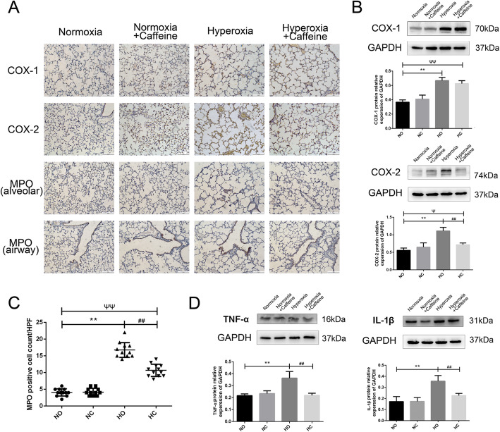
Fig. 4.
Caffeine reduced the inflammatory reaction in neonatal mouse lung. Hyperoxia exposure could increase the expression of COX-1, COX-2, MPO, IL-1β and TNF-α in the lung, as shown, and caffeine treatment could decrease the levels of COX-2, MPO, IL-1β and TNF-α. a The expression of COX-1, COX-2 and MPO protein in lung as determined by IHC staining (light microscopy, × 100). Scale bars = 100 μm. b The protein levels of COX-1 and COX-2 in the lung as determined by western blotting. c The number of MPO-positive cells in the lung alveolar space in a high-power field (HPF). d The protein levels of IL-1β and TNF-α in the lung as determined by western blotting. The values are the mean ± SD; n = 6 mice/group. **P < 0.01 Hyperoxia (HO) group versus Normoxia (NO) group, ##P < 0.01 Hyperoxia + Caffeine (HC) group versus HO group, ΨP < 0.05, ΨΨP < 0.01 NO group versus HC group

