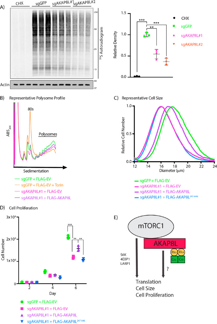
Figure 4.
AKAP8L regulates protein translation, cell size, and proliferation. A, depletion of AKAP8L decreases global protein synthesis. AKAP8L KO cells were incubated in methionine and cysteine-free Dulbecco's modified Eagle's medium for 1 h. Cycloheximide (CHX) treatment for 1 h was used as a positive control. 35S-labeled l-methionine and l-cysteine mix was then added to the medium for 10 min, and newly synthesized proteins were detected by autoradiography. Values are displayed as means ± S.D. (error bars). Significance was analyzed using Student's t test. B, loss of AKAP8L reduces the number of polysomes. AKAP8L KO cells with or without stably expressing full-length FLAG-tagged AKAP8L were subjected to polysome profiling. Negative control WT cells were treated with 100 nm Torin1 for 1 h. A representative image is shown from three biological replicates. C, loss of AKAP8L reduces cell size. The size of AKAP8L KO cells with or without stably expressing full-length FLAG-tagged AKAP8L or FLAG-tagged AKAP8L 247–646 was measured using a Coulter counter. Samples with the closest value to the mean are plotted as a representative image. Significance (p) was as follows: sgGFP versus sgAKAP8L#1 + FLAGEV, <0.01; sgGFP versus FLAGAKAP8L 247–646, <0.01; sgAKAP8L#1 + FLAGAKAP8L versus sgAKAP8L#1 + FLAGEV, <0.001; sgAKAP8L#1 + FLAGAKAP8L versus sgAKAP8L#1 + FLAGAKAP8L 247–646, <0.001. Significance was analyzed using Student's t test. The number of biological repeats is n ≥ 3. The number of technical repeats of each sample is n = 3 per experiment. D, loss of AKAP8L reduces cell proliferation. AKAP8L KO cells with or without stably expressing full-length FLAG-tagged AKAP8L or FLAG-tagged AKAP8L 247–646 were counted on the indicated days using trypan blue and a Bio-Rad automated cell counter. Values are displayed as means ± S.D. Significance was analyzed using Student's t test. The number of biological repeats is n ≥ 3. The number of technical repeats of each sample is n = 3 per experiment. E, working model of AKAP8L regulating translation, cell size, and proliferation. Significance is indicated as follows. **, p < 0.01; ***, p < 0.001.

