Figure 1.
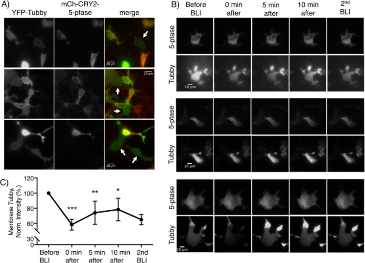
BLI-induced 5-ptase recruitment leads to translocation of the Tubby PIP2-binding domain from the membrane to cytoplasm. A, three representative fluorescence micrographs of HEK 293 cells transfected with YFP-Tubby (left), CIBN and mCh-CRY2–5-ptase (middle) after BLI. The merged images are shown to the right, with white arrows indicating cells expressing only YFP-Tubby. B, three representative cells imaged under TIRF microscopy showing mCh-CRY2–5-ptase (top panels) and YFP-Tubby (bottom panels) before and 0, 5, and 10 min after BLI, then immediately following a second BLI. C, summary graph of membrane YFP-Tubby normalized to the TIRF fluorescence intensity of membrane YFP-Tubby before BLI. Data represent a mean ± S.E. of n = 5 or 6 cells of n = 2 independent experiments; ***, p < 0.0001; **, p < 0.005; *, p < 0.05 compared with the cells before BLI.

