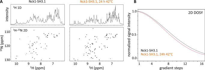
Figure 10.
NMR shows structured Nck1-SH3.1 species in solution. A, the 1H (top) and 15N HSQC (bottom) spectra of the Nck1-SH3.1 dimer (left panel) show significant differences compared with spectra obtained from the identical sample after gentle heat treatment (24 h, 42 °C; right panel). Conformational differences in the two Nck1-SH3.1 species are reflected by numerous chemical shift perturbations of backbone (NH) resonance signals. Note the decreased line widths after heat treatment, indicating an increased rotational correlation time due to formation of monomers. The overall signal dispersion is not affected; thus, both species adopt stable structures, and no aggregation is present. B, DOSY detects differences in diffusion rates consistent with a roughly 2-fold mass difference. The faster signal decay for the heat-treated Nck1-SH3.1 indicates increased average diffusion rates for the molecules in this sample.

