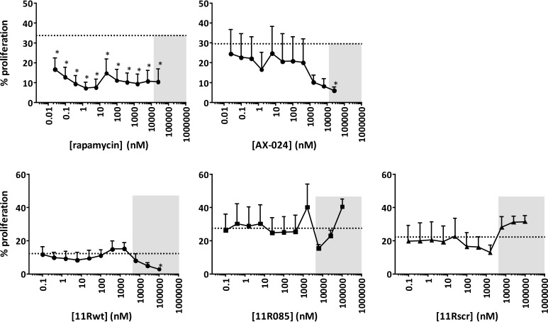
Figure 2.
CD3ϵ-derived peptides do not inhibit T cell proliferation. CD4 T cell proliferation was assessed upon stimulation with anti-CD3ϵ by CFSE dilution as a measure for cellular proliferation, analyzed on day 4. For rapamycin and AX-024–treated cells, the dashed line indicates control proliferation at a DMSO concentration of 0.2%. For CD3ϵ-derived peptides 11Rwt, 11R085, and 11Rscr, the dashed line indicates control proliferation at 0.3% DMSO, 0.3% PBS. 11Rwt has sequence R11-G3-RGYNKERPPPVPNPDY, 11R085 R11-G3-Q(dK)KECPPPVPKRDY, and 11Rscr R11-G3-PKVRECPDYK(dK)PQP. A polyarginine tag is used to enable cell penetration of the peptides (10). The gray boxes indicate concentrations of compound leading to decreased cell viability. These values should be disregarded. Binding of peptides 11Rwt and 11R085, but not 11Rscr, to Nck-SH3.1 was confirmed by NMR spectroscopy (Fig. S1). Asterisks, p < 0.05 as determined by a repeated-measure one-way analysis of variance with Holm–Sidak's multiple-comparison test to DMSO/PBS. Dotted lines, mean DMSO/PBS values. Error bars, S.D.

