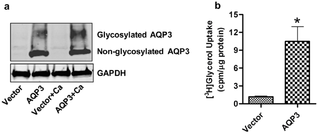
Figure 1. Validation of expression and activity of re-expressed AQP3 in AQP3-knockout keratinocytes.

Primary cultures of AQP3-knockout keratinocytes were allowed to reach approximately 70-80% confluence and then infected with adenoviruses expressing either wild-type AQP3 or vector alone using a MOI of 25 for 24 hours. Fresh control medium (50μM calcium) or medium containing 125μM calcium was added for another 24 hours. (a) Total cell lysates were analyzed by Western blotting using antibodies against AQP3 and the GAPDH loading control. AQP3 is seen as a non-glycosylated, 28 kDa band and a diffuse band at 35-40 kDa, representing the glycosylated form. The figure shown is representative of three independent experiments. (b) [3H]glycerol uptake by the cells is shown as cpm/μg protein. The data represent the means ± SEM from three independent experiments. *p<0.05 versus vector-infected keratinocytes.
