
Figure 4. The PLD2 inhibitor, CAY10594, inhibited the AQP3 re-expression-induced increase in mRNA levels of markers of keratinocyte differentiation.
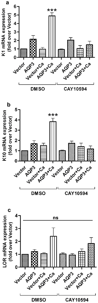
Primary AQP3-knockout mouse keratinocytes were allowed to reach approximately 70-80% confluence and then infected with adenoviruses expressing either wild-type AQP3 or vector alone using an MOI of 25 for 24 hours. Medium containing the PLD2-selective inhibitor, CAY10594 (1 μM) alone or in combination with an elevated calcium concentration (125μM) was added to the keratinocytes for an additional 24 hours. (a) The cells were harvested for qRT-PCR analysis, and mRNA expression of (a) keratin 1 (K1), (b) keratin 10 (K10) and (c) loricrin (LOR) was determined using the ΔΔCt method with GAPDH as the endogenous control. The results are shown as the fold over the vector-infected group and were derived from at least three independent experiments. *** p<0.001 compared to all other groups; ns, non-significant.