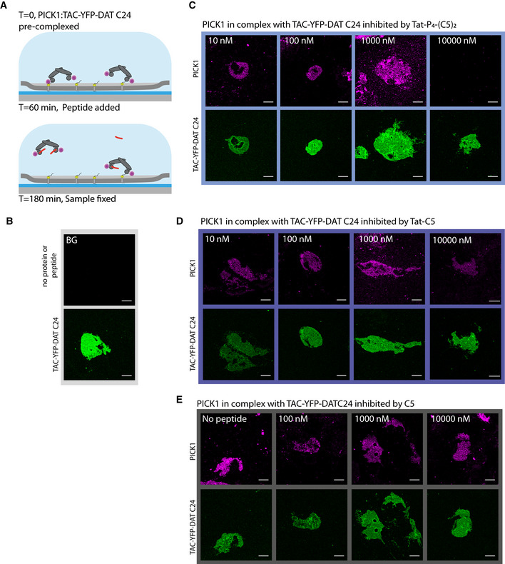
Representative fluorescence confocal images of SCMS expressing TAC‐YFP‐DAT C24 (green) incubated with fluorescently labeled PICK1 (magenta) and subsequently incubated with unlabeled peptide at indicated concentrations. Visible reduction in binding of PICK1 (magenta) is observed at 10 μM of Tat‐P
4‐(C5)
2, but not for Tat‐C5 and C5, suggesting that Tat‐P
4‐(C5)
2 can facilitate dissociation of PICK1 from SCMSs. For curves in Fig
1F, images were pooled from three independent experiments. Note that images with 10,000 nM peptide are the same as used in Fig
2E.

