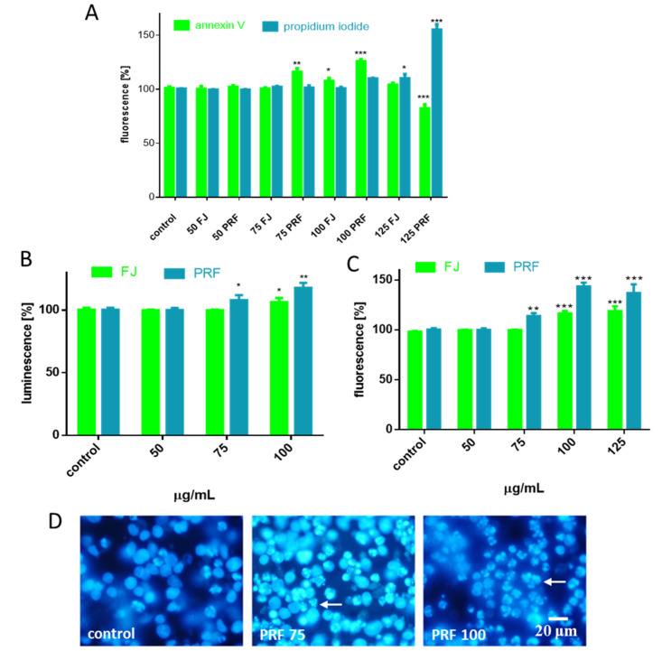
Figure 4.
The influence of 24 h of exposure of FJ and PRF on phosphatidylserine externalization on the outer membrane leaflet of apoptotic MIN6 cells and membrane permeabilization detected with Annexin-V-FITC assay kit and propidium iodide staining (A); extract influence on caspase-9 activation (B) and caspases-3/7 activation (C); DAPI-stained nuclei and chromatin condensation observed using fluorescence microscope (Olympus CKX41, Japan), 400 × magnification (D). Control cells were not exposed to any compound but the vehicle; values are means ± standard deviations from at least three independent experiments, n ≥ 9; statistical significance was calculated versus control cells (untreated), * p ≤ 0.05, ** p ≤ 0.01, *** p ≤ 0.001.

