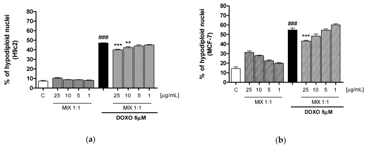
Figure 5.
Apoptosis detection by propidium iodide (PI) staining of hypodiploid nuclei after H9c2 (a) and MCF-7 (b) incubation with MIX 1:1 ratio (1–25 μg/mL), alone and in combination with DOXO (5 μM) for 24 h. Data are expressed as mean ± s.e.m. of % of hypodiploid nuclei. *** and ** denote respectively p < 0.001 and p < 0.01 vs DOXO; ### denotes p < 0.001 vs C.

