Figure 4.
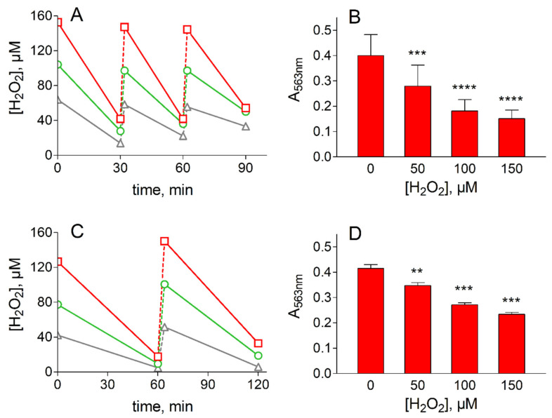
Treatment of lens epithelial cells with H2O2. (A) and (B) refer to H2O2 removal and viability assay in HLEC, respectively. (C) and (D) refer to H2O2 removal and viability assay in bovine lens epithelial cells (BLEC), respectively. Panel (A): HLEC at 70% confluence (2.5 × 10⁶ cells) were incubated in Minimum Essential Medium (7 mL) at 37 °C; at the indicated times cells were supplemented with 50 (gray), 100 (green) or 150 (red) µM H2O2 and the concentration of the peroxide was measured as described in Section 2.4. C: BLEC at 70% confluence (1.0 × 10⁶ cells) were incubated in HBSS (7 mL) at 37 °C; at the indicated times, cells were supplemented with 50 (gray), 100 (green) or 150 (red) µM H2O2 and the concentration of the peroxide was measured as described in Section 2.4. (C) and (D): Cell viability was evaluated by MTT assay (see Section 2.3) at the end of overall oxidative treatment at the indicated concentrations of H2O2. The values represent the mean ± SEM of at least three independent experiments. Significance was evaluated with respect to untreated cells (** p < 0.01, *** p < 0.001, **** p < 0.0001).

