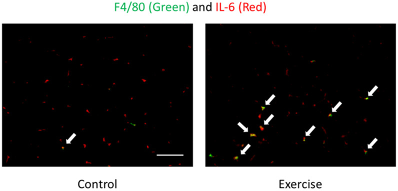
Figure 1.
Localization of F4/80 (macrophages) (green) and IL-6 (red) of skeletal muscle after exercise detected by immunofluorescence staining [61]. Arrows (yellow) indicate F4/80 and IL-6 double positive cells. The signals of IL-6 were mainly observed in the interstitial space. Exercise increased F4/80 and IL-6 double positive cells but not IL-6 positive myocytes. This result suggests macrophages are one of the sources of IL-6.

