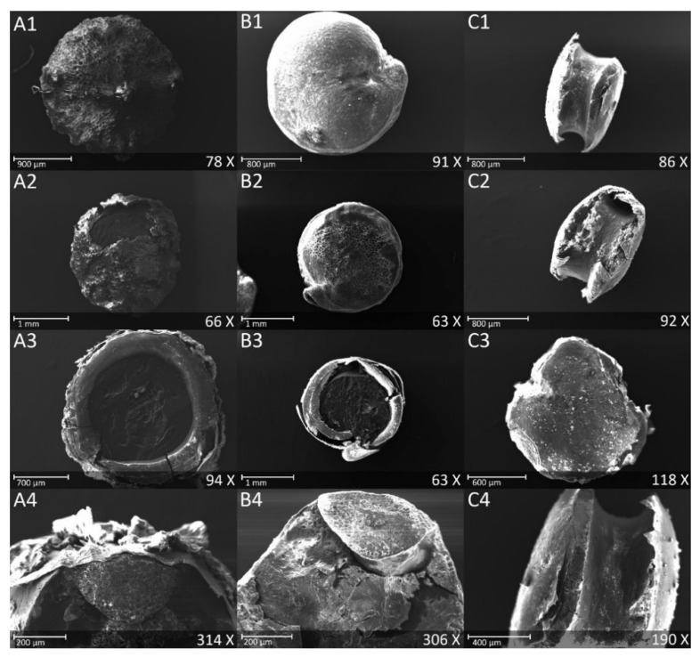
Figure 2.
Scanning Electron Microscope images of quinoa seeds cv Titicaca. (A) Raw seeds: (1) Dorsal surface, (2) Ventral surface, (3) Cross section showing different seed layers, (4) Longitudinal section showing the embryo. (B) Polished seeds: (1) Dorsal surface, (2) Ventral surface with residual outer layer, (3) Cross section showing the embryo and the seed layers, (4) Longitudinal section showing the embryo. (C) Semolina: (1) Lateral view, (2) Lateral view showing residual layers and (3) Dorsal surface, (4) Lateral view showing the empty location of the embryo.

