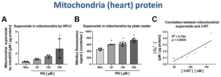
Figure 7.
Detection of superoxide generation by mitochondria by mitoSOX HPLC and correlation with 3-NT levels. Isolated heart mitochondria were nitrated by PN at concentrations of 50–250 µM. These samples were split, one aliquot was used for measurement of mitochondrial superoxide formation using mitoSOX/HPLC and the other aliquot for determination of free 3-NT levels by HPLC/ECD after digestion. Mitochondrial superoxide formation was determined using HPLC-based quantification of 2-OH-mito-E+ (A) and ROS formation was measured using a fluorescence plate reader assay for the mitoSOX oxidation products (B). The yield of mitochondrial 3-NT was correlated with superoxide formation rate for the different PN-treated samples (C). HPLC/ECD analysis was performed with 20 µL of sample at 27 °C with isocratic elution (1 mL/min, mobile phase: 26.3 mM sodium citrate and 10.9 mM sodium at pH 3.75 with 3.5 v/v% methanol; 3-NT eluted at 4.05 min). Data are presented as mean ± SD of n = 3–5 (A); n = 4 (B) and n = 8 (C) independent experiments. * indicates p < 0.05 versus Mito untreated control group.

