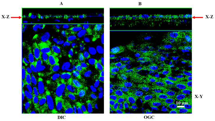
Figure 8.
Localization of DIC and OGC in polarized RPE monolayers. RPE monolayers grown on Transwell filters were stained for OGC and DIC. The specimen was viewed on an LSM 770 laser-scanning microscope. (A) immunofluorescence staining of DIC in RPE monolayers showing predominant apical staining (X–Z plane). However, OGC has a wide distribution, expressed both in the apical as well as basolateral domains (B).

