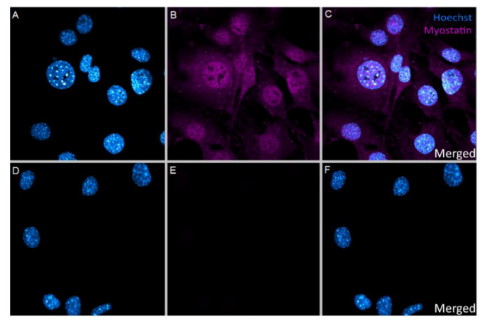
Figure 1.
(A–F): Immunocytochemical staining of VSMCs. (A–C)—VSMCs treated with 10 nM myostatin showed uptake of myostatin in the cell, concentrating mostly in the nucleus. (A)—Hoechst only, (B)—Myostatin only, (C)—Merged image. (D–F)—VSMCs not treated with myostatin showed no endogenous myostatin presence in the cells at all. (D)—Hoechst only, (E)—Myostatin only, (F)—Merged image. Picture taken with 63× magnification.

