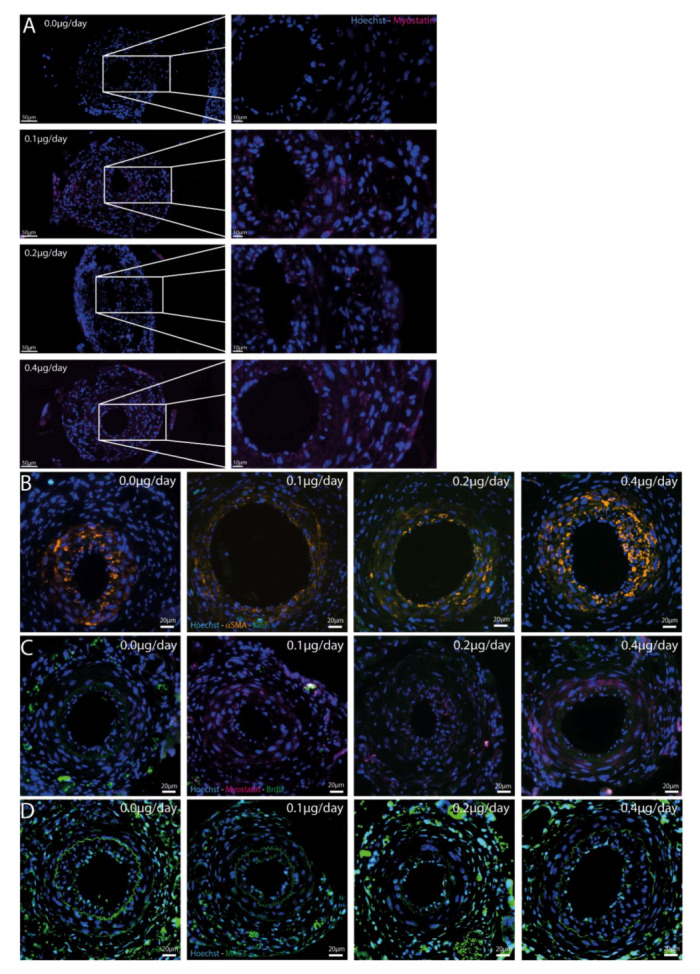
Figure 5.

(A–D): Immunofluorescent stainings in different treatment groups. (A)—Myostatin-Hoechst immunofluorescent double staining. Representative mouse of every treatment group is shown and zoom-in of femoral artery shows presence of myostatin in all treated groups, but not in the control group. (B)—Ki-67-αSMA-Hoechst immunofluorescent triple staining in cuffed femoral arteries of different treatment groups shows few proliferating cells, especially not in the αSMA area. (C)—BrdU-Myostatin-Hoechst immunofluorescent triple staining in cuffed femoral arteries of different treatment groups shows again little proliferating cells and those cells are not double stained with myostatin. (D)—Hoechst-MAC3 immunofluorescent double staining of cuffed femoral arteries showed macrophages in all groups in both the intimal and medial layers, but no differences were found.
