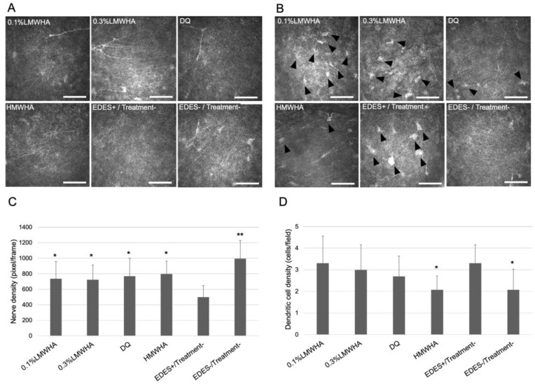
Figure 5.
Comparison of subepithelial nerve density and presumed dendritic cell density measured by in vivo confocal microscopy. (A) Representative confocal microscopy image showed marked decrease of subepithelial nerve in the EDES+/Treatment− group. (B) Representative confocal microscopy image showed marked increase of subepithelial dendritic cell infiltration in the EDES+/Treatment− group. (C) The mean subepithelial nerve density in the EDES+/Treatment− group was significantly lower than other groups. (D) The mean subepithelial dendritic cell density in the HMWHA and EDES−/Treatment− groups was significantly lower than the EDES+/Treatment− group. * and ** represent p < 0.05 and p < 0.001 when each group was compared with the EDES+/Treatment− group, respectively. Scale bar shows 100 µm. Black arrowheads show presumed dendritic cells. LMWHA, low molecular weight hyaluronic acid (HA); DQ, diquafosol sodium; HMWHA, high molecular weight HA; EDES, environmental dry eye stress.

