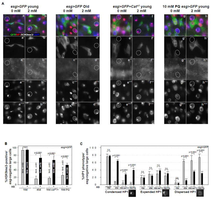
Figure 3.
β-HB inhibits age- and oxidative stress-induced heterochromatin instability in midgut ECs. (A) Guts from 10-day-old esg > GFP flies (a–b”’’), 45-day-old esg > GFP flies (c–d”’’), and 10-day-old esg > GFP + Catn1 mutant flies (e–f’’”), without (a–a’’”, c–c’’’’, e–e’’’’) or with (b–b’’’’, d–d’’’’, f–f’’’’) 2 mM β-HB feeding for seven days, were stained with anti-HP1 (red), anti-H3K9me3 (white), anti-GFP (green), and DAPI (blue). Ten-day-old esg > GFP flies, without (g–g’’”) or with (h–h’’”) 2 mM β-HB feeding for six days, were treated with 10 mM PQ in standard media for 20 h, after which their guts were stained with anti-HP1 (red), anti-H3K9me3 (white), anti-GFP (green), and DAPI (blue). a’, b’, c’, d’, e’, f’, g’, and h’ indicate enlarged GFP stained images. a”, b”, c”, d”, e”, f”, g”, and h” indicate enlarged H3K9me3 stained images. a”’, b’”, c’”, d’”, e’”, f’”, g’”, and h’” indicate enlarged HP1 stained images. a”’’, b’’’’, c’’”, d’’”, e’’”, f’’”, g’’”, and h’’” indicate enlarged DAPI stained images. White dotted circles indicate the nuclei of ECs (esg-negative cell). Original magnification is 400×. (B) Graph showing the proportion of H3K9me3-positive cells in GFP-negative large cells (ECs) in 10-day-old esg > GFP, 45-day-old esg > GFP, 10-day-old esg > GFP + Catn1 mutant, and 10-day-old PQ-treated esg > GFP flies, with (black bar) or without (gray bar) β-HB feeding for seven days. (C) Graph showing the proportion of condensed, expanded, and dispersed HP1 phenotype in GFP-negative large cells in 10-day-old esg > GFP, 45-day-old esg > GFP, 10-day-old esg > GFP + Catn1 mutant, and 10-day-old PQ-treated esg > GFP flies, with (black bar) or without (gray bar) β-HB feeding for seven days. N is the number of observed guts, and n is the number of observed cells. The error bar represents standard error. p-values were calculated using Student’s t-test. n.s. indicates not significant (p > 0.05).

